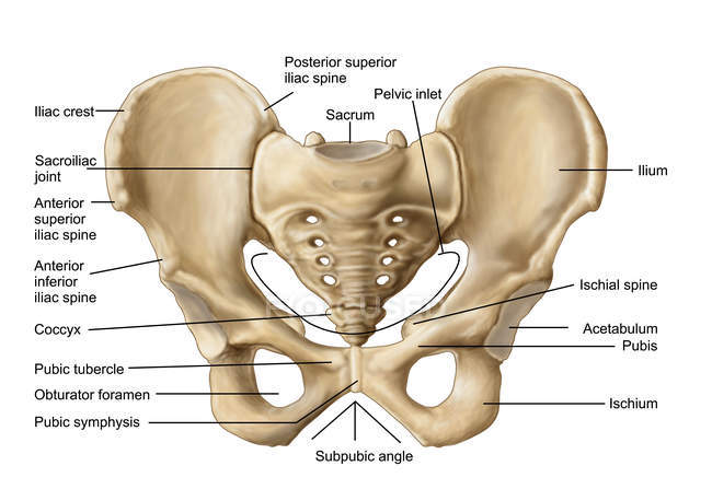
The pelvis consists of two hip bones attached at the front anterior by the pubic symphysis and at the back posterior by the sacrum. The lumbosacral joint is a symphysis secondary cartilaginous joint between the fifth lumbar vertebra and the base of the sacrum.
Anatomy Of Human Pelvic Bone With Labels
Browse Stock Photos Vectors and Much More.

. Follow the directions and lists to label the structures you are to learn on the appropriate figures. Mandible light green temporal bone dark blue. View the full answer.
Articulated Lower limb 1. The figure below is a lateral view of the head. There is a printable worksheet available for download here so you can take the quiz with pen and paper.
Label the bone features bone markings of the radius and ulna anterior and posterior views by clicking and dragging the labels to the correct location. Click on the tags below to find other quizzes on the same subject. About this Quiz.
The ovary is connected to the lateral pelvic wall with the suspensory ligament of the ovary. Most but not all features you are required to know are shown on the following pages. It is bounded laterally by the iliac bones posteriorly by the lumbar spine and anteriorly by the abdominal wall.
Maxilla yellow parietal bone light blue. Label the following structures on the lateral view of the pelvis below. Acoustic Meatus References The temporalis muscle passos modial to this structure.
Weve got two hip bones a sacrum and a coccyx. The false pelvis forms the lower part of the abdominal cavity. 2 in the curve of Carras.
Anatomy and Physiology questions and answers. The canal of the true pelvis is bent forward in its lower portion see Fig. True and False Pelvis Lesser and Greater Pelvis The pelvis is separated into two regions.
Bony Landmarks of the Pelvis and Thigh by Iron-Butterfly 385 plays 22p Image Quiz. Subscribe and Save Now. Study from the bone list or your textbook after you marked the drawings as instructed on page 6-2.
Its also called the greater pelvis. This is an online quiz called Labeling the Bones of the Skull. Youve got the upper region the superior part of the pelvic bone which is called the false pelvis.
Sphenoid bone Mastoid process Occiptial Condyle. Label figures 171 and 172. Simple Licensing Dedicated Customer Support.
Round ligament of the uterus uterine tube ovarian ligament ureter uterosacral ligament. Label the bones and anatomical features of the inferior view of the skull 16 Parietal hone Zygomatic arch Mastoid notch eBook Condylar canal Ext. Start studying Anatomy 2017- Unit 3 Label the Bones of the Pelvic Girdle Anterior view.
On the line below each view label. Label the posterior surface of the right scapula by clicking and dragging the terms to the correct location. Ad Try Now and Get 10 Free Photos.
Learn vocabulary terms and more with flashcards games and other study tools. After you have labeled the bones coloring them using the following chart. Learn vocabulary terms and more with flashcards games and other study tools.
BONES OF THE AXIAL AND APPENDICULAR SKELETON. Pelvis cavity medial and lateral view by agrant12 551 plays 11p Image Quiz. Start studying Lab 17.
Posterior inferior iliac spine anterior superior iliac spine superior pubic ramus anterior gluteal line Solutionpdf Experts Answer. Learn vocabulary terms and more with flashcards games and other study tools. Start studying pelvic girdle label.
Label the following bones. 1 ANATOMY WORKBOOK BY. AP291 by shimes 598 plays 14p Image Quiz.
Nasal bone purple occipital bone dark green. Ann Stoeckmann Bones of the Pelvic Girdle and Lower Limb The figure titles on the separate document are listed below. Os means bone and coxae means of the hip so its the bone of the hip the hip bone.
Each hip bone consists of an ilium ischium and pubis. Lable the pelvis by USSUMW_Hayley 483 plays 11p Image Quiz. The bones of the pelvis articulate with each other via four joints.
Superior view of female pelvis and surrounding endopelvic fascia. Canine pelvis x-ray 3 by GracynVH 387 plays 15p Image Quiz. Do not spend your.
Posterior to anterior these are the lumbosacral sacroiliac sacrococcygeal hip and pubic symphysis joints. Start studying Pelvis labeling. Label the structures of the pelvis.
94 16 ratings Answer The label is indicated from RIGHT SIDE of the image to LEFT SIDE TOP to BOTTOM Right side 3 label Ilium - It i. Label color palpate and answer the questions listed for each figure. This is an online quiz called Label the Pelvis.
After you have studied the bones in lab label the drawings as a self-test. This quiz has tags. Structures shown on the lateral side of the uterus from anterior to posterior.
It has little obstetrical significance. Learn vocabulary terms and more with flashcards games and other study tools. Innominate ilium iliac crest anterior superior iliac spine posterior superior iliac spine greater sciatic notch portion in ischium iliac fossa ischium ischial tuberosity ischial spine lesser sciatic notch pubis pubic symphysis joint between pubic bones pubic.
Examine the bones of the pelvic girdle and locate the following. Intervertebral disc Lumbar vertebra Sacrum Coccyx Piibis Ischium. Use code PICK10FREE at checkout.
There is a printable worksheet available for download here so you can take the quiz with pen and paper.
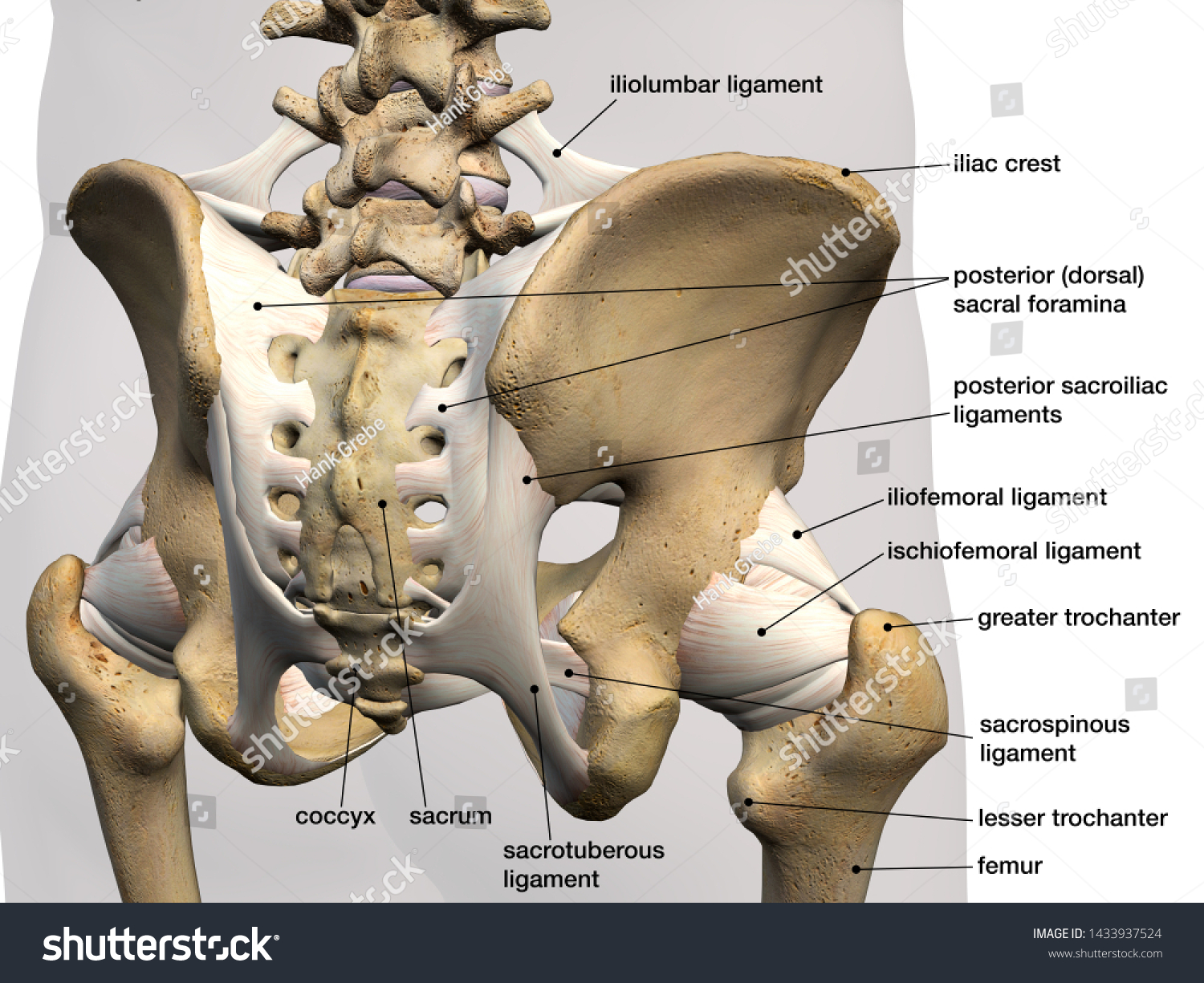
Pelvic Hip Bones Ligaments Labeled Posterior Stock Illustration 1433937524
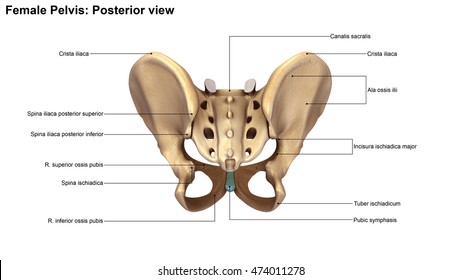
Skeleton Pelvis Posterior View 3d Illustration Stock Illustration 474011278

The Pelvic Girdle And Pelvis Anatomy And Physiology I

Coxal Pelvic Bone Posterior View With Labels Appendicular Skeleton Visual Atlas Page 18 Anatomy Flashcards Medical Anatomy Pelvic Bone

Lab 17 Figure 17 1 Pelvis Diagram Quizlet

Anatomy 2017 Unit 3 Label The Bones Of The Pelvic Girdle Anterior View Diagram Quizlet
Human Skeleton System Pelvis With Labels Anatomy Stock Photo Download Image Now Istock

8 3 The Pelvic Girdle And Pelvis Anatomy Physiology
0 comments
Post a Comment